Microscopic anatomy and organization of skeletal muscle exercise 11 delve into the intricate structure and function of skeletal muscle, exploring how exercise influences its adaptations. This detailed examination unravels the mechanisms underlying muscle growth, metabolism, and force production, providing a comprehensive understanding of skeletal muscle physiology.
From the microscopic architecture of myofibrils to the dynamic adaptations induced by exercise, this topic unveils the fascinating interplay between muscle structure and function. Understanding these concepts is crucial for optimizing training programs, enhancing athletic performance, and maintaining muscle health.
Microscopic Anatomy of Skeletal Muscle

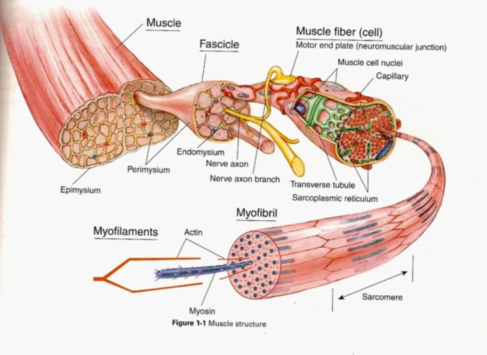
Skeletal muscle tissue is composed of elongated, cylindrical muscle fibers. Each muscle fiber is a single, multinucleated cell that contains numerous myofibrils, which are the contractile units of muscle.
Myofibrils, Myofilaments, and Sarcomeres
Myofibrils are composed of repeating units called sarcomeres, which are the basic units of muscle contraction. Sarcomeres are bounded by Z-disks and contain two types of myofilaments: thick filaments (myosin) and thin filaments (actin).
Connective Tissue in Muscle Organization
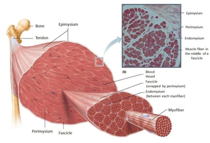
Connective tissue surrounds and supports muscle fibers and bundles them into fascicles. The outermost layer of connective tissue, called the epimysium, surrounds the entire muscle. The perimysium surrounds each fascicle, and the endomysium surrounds each individual muscle fiber.
Exercise-Induced Adaptations in Skeletal Muscle: Microscopic Anatomy And Organization Of Skeletal Muscle Exercise 11
Muscle Fiber Size and Type, Microscopic anatomy and organization of skeletal muscle exercise 11
Exercise can increase muscle fiber size (hypertrophy) and change the proportion of different fiber types. Type II fibers, which are fast-twitch and glycolytic, are more responsive to hypertrophy than Type I fibers, which are slow-twitch and oxidative.
Molecular Mechanisms of Muscle Hypertrophy and Hyperplasia
Muscle hypertrophy is primarily driven by increased protein synthesis and decreased protein degradation. Exercise activates signaling pathways that stimulate protein synthesis and inhibit protein degradation.
Role of Satellite Cells in Muscle Adaptation
Satellite cells are muscle stem cells that can differentiate into new muscle fibers. Exercise stimulates satellite cell activation and proliferation, contributing to muscle growth and repair.
Microcirculation and Oxygen Delivery

Microcirculation of Skeletal Muscle
The microcirculation of skeletal muscle consists of capillaries, arterioles, and venules. Capillaries are the smallest blood vessels and allow for the exchange of oxygen and nutrients between the blood and muscle cells.
Structure and Function of Capillaries, Arterioles, and Venules
Capillaries are thin-walled vessels that allow for the diffusion of oxygen and nutrients. Arterioles regulate blood flow to capillaries, and venules collect blood from capillaries and return it to the heart.
Factors Influencing Blood Flow to Skeletal Muscle during Exercise
Blood flow to skeletal muscle is increased during exercise to meet the increased oxygen and nutrient demands. Factors that influence blood flow include muscle fiber type, exercise intensity, and local temperature.
Metabolism and Energy Production

Metabolic Pathways Used by Skeletal Muscle during Exercise
Skeletal muscle uses several metabolic pathways to produce energy during exercise, including aerobic metabolism (oxidative phosphorylation) and anaerobic metabolism (glycolysis and lactic acid fermentation).
Role of Glucose, Fatty Acids, and Other Substrates in Energy Production
Glucose is the primary fuel for skeletal muscle during high-intensity exercise, while fatty acids are the primary fuel during low-intensity exercise. Other substrates, such as ketones and amino acids, can also be used for energy production.
Regulation of Muscle Metabolism during Exercise
Muscle metabolism is regulated by several factors, including exercise intensity, muscle fiber type, and hormonal signals. Exercise intensity primarily determines the balance between aerobic and anaerobic metabolism.
Neuromuscular Junction and Motor Unit Recruitment

Structure and Function of the Neuromuscular Junction
The neuromuscular junction is the site of communication between a motor neuron and a muscle fiber. The motor neuron releases acetylcholine, which binds to receptors on the muscle fiber, triggering muscle contraction.
Process of Motor Unit Recruitment and Its Role in Muscle Force Production
Motor units are groups of muscle fibers that are innervated by a single motor neuron. Motor units are recruited in an orderly fashion during muscle contraction, with smaller motor units being recruited first.
Factors Influencing Motor Unit Recruitment during Exercise
Motor unit recruitment is influenced by several factors, including exercise intensity, muscle fiber type, and the level of fatigue. Higher exercise intensities and muscle fiber types recruit more motor units.
FAQ
What are the key components of skeletal muscle tissue?
Skeletal muscle tissue consists of myofibrils, which are composed of myofilaments (actin and myosin) arranged in repeating units called sarcomeres.
How does exercise affect muscle fiber size and type?
Exercise can increase muscle fiber size (hypertrophy) and shift fiber type composition towards more fatigue-resistant fibers (Type I).
What is the role of satellite cells in muscle adaptation?
Satellite cells are muscle stem cells that contribute to muscle growth and repair.
How does blood flow to skeletal muscle change during exercise?
During exercise, blood flow to skeletal muscle increases to deliver oxygen and nutrients.